

Important bodily activities are carried out by human lungs. They are essential because they enable you to speak and smell, as well as allow warm air to moisten it to the appropriate level of humidity for your body.
There are occasions when the lungs malfunction, are damaged, or are exposed to diseases. Breathlessness and pain are the most typical symptoms of this.
Lung tissues, usually the cells that line the airways, can develop cancer, which is known as lung cancer. Men and women who develop cancer die mostly as a result of it.
While a pleural effusion is fluid in the normally air-filled gaps in the lungs, fluid on an X-ray is white, air is black; therefore a portion of the lung is white instead of black, lung cancer is typically a white lump in the air containing thus black lung on the chest x-ray.
Lungs need to be kept safe and regular in function through a healthy approach to lifestyle, which would include keeping weight in check, regular exercise, a nutritious diet, and no smoking.
Keep reading to know more about human lungs, their function, and the harmful occurrences of injury and cancer around them.
What Is A Lung?
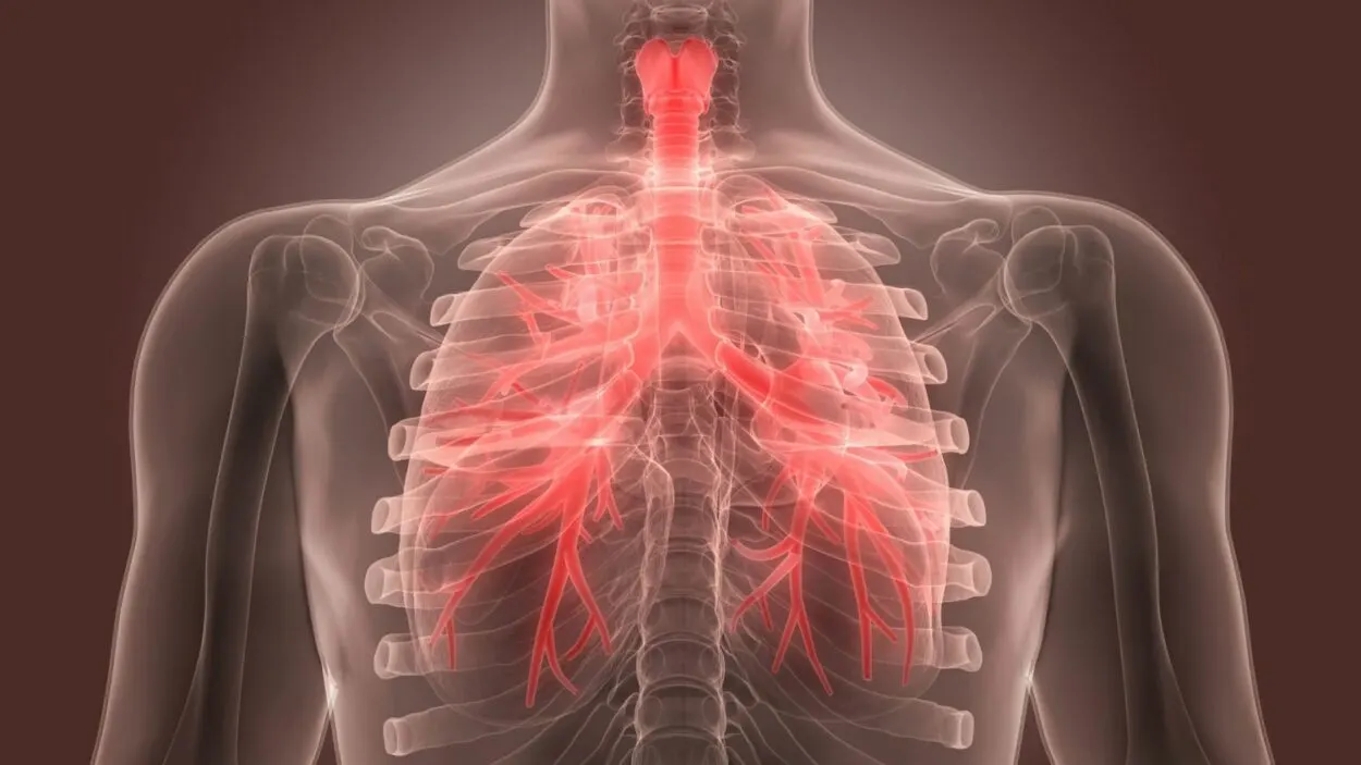
The respiratory system, a collection of organs and tissues that cooperate to help you breathe, includes your lungs. The respiratory system’s main jobs are to move oxygen throughout the body and get rid of extra carbon dioxide.
Within your chest cavity, your lungs are located on either side of your heart. They are the primary respiratory system organs.
The left lung is divided into two lobes, whereas the right lung has three lobes (parts).
The spongy texture of the lung is a result of the alveoli. The inside of these alveoli is lined by pneumocytes, flattened epithelial cells with a single entrance.
Three types of tissue make up the alveolar wall or septum: surface epithelium, supporting tissue, and a vast network of continuous capillaries.
The common functions of the lungs from many are as follows:
| Lungs Function | Description |
| Mouth and Nose | It enables you to talk and smell. |
| Humidity Maintenance | It raises the temperature of the air to that of your body and adds moisture to it to the proper degree of humidity. |
| Oxygen Regulation | It provides your body’s cells with oxygen. |
| Removal of Waste | When you exhale, waste gases from your body, including carbon dioxide, are removed. |
Common Disorders Affecting Lungs


The color of healthy lungs is pinkish-gray. You’ve probably seen images that contrast the lungs of smokers with those of non-smokers.
Damaged lungs have a deeper grey color and may have black patches. These harms may manifest as sudden accidents, illness, cancer, or infections.
There are several distinct lung diseases. While some are minor and transient, others are more serious and chronic. Let’s look at them now.
| Lung Disease Name | Description |
| Asbestosis | Your lungs and pleural tissue become scarred after inhaling asbestos fibres. |
| Asthma | Breathing becomes challenging when airways are constricted. |
| Bronchitis | Coughing is this condition’s primary symptom. You can have acute or chronic bronchitis. |
| Cystic Fibrosis | Your lungs and other organs develop an accumulation of sticky mucus as a result of this genetic illness. |
| Influenza | The flu is a lung condition brought on by a virus. |
| Lung Cancer | Smoking cigarettes is a major risk factor for lung cancer. |
What Is A Lung Cancer?
Usually, in the cells that line the airways, lung tissues can become cancerous and produce lung cancer.
It is the primary factor in both men’s and women’s cancer-related deaths. The two primary types are small-cell lung cancer and non-small-cell lung cancer.
The leading cause of lung cancer risk is cigarette smoking; about 80% to 90% of lung cancer deaths in the US are attributed to smoking cigarettes.
Lung cancer risk is increased by using tobacco products, including cigars, pipes, and other cigarettes.
The second most common cause of lung cancer is radon exposure; a radioactive gas called radon, which has no color or smell, naturally occurs in soil.
One’s risk of acquiring lung cancer may also be influenced by genetic factors; you may have an increased risk of developing lung cancer if your family has a history of the disease.
Some common symptoms of lung cancer are:
- Coughing
- Chest pain
- Shortness of breath
- Wheezing
- Coughing up blood
- Feeling very tired all the time
Lung Cancer On An Xray


Low-dose computed tomography, often known as an LDCT scan or low-dose CT, is the only screening procedure for lung cancer that is advised.
During an LDCT scan, which is painless and only takes a few minutes, an X-ray scanner uses a low dosage (amount) of radiation to get exact pictures of your lungs while you are reclining on a table.
Typically, the first test performed to identify lung cancer is a chest X-ray. On X-rays, the majority of lung tumors show up as a white-gray mass.
Lung cancer cells can occasionally be visible when sputum is coughed up and studied under a microscope; this is particularly true if you have a cough and are generating sputum.
What Is Pneumonia Pleural Effusion In Lungs?
When fluid accumulates between the lung and the chest wall, a pleural effusion happens. Pneumonia and consequences from heart, liver, or kidney illness are only a few causes of this.
Pleural effusion, a potentially hazardous condition that can appear to be something less concerning, is fluid around the lung.
What can appear to be coughing or chest pain brought on by a nasty cold could actually have detrimental effects on one’s health. It’s also not all that uncommon.
Each year, pleural effusion is identified in more than 1.5 million people in the US.
Pleural effusion symptoms can be none, shortness of breath, coughing, and more. The more fluid that accumulates, the more probable it is that symptoms may become apparent.
Pleural Effusion On Xray


The detection of pleural effusion is quite sensitive with upright chest radiography. The most accurate radiographic pictures for identifying free pleural effusion are lateral decubitus projections.
It is possible for even big, loculated, or unusual effusions to show significant gravitational movement that reveals their nature.
An effusion is a fluid in a rather open area, so as you change positions, it usually moves due to gravity.
Blunting of the costophrenic angle is another most common but least reliable indicator of pleural effusions on supine radiographs.
Loss of the hemidiaphragm and an increase in hemithorax density are two additional indicators that can be helpful. A pleural effusion is not ruled out by a typical supine radiograph.
Difference Between Pneumonia, Pleural Effusion, And Lung Cancer On An X-Ray
As distinct pathologic processes, lung cancer, pleural effusion, and pneumonia each have unique radiographic manifestations.
All three result in more white spots on the X-ray because cells, pus, or fluid occupy the spaces that should have been filled with air.
Despite the fact that both pneumonia and lung cancer frequently replace air space in the lungs, their patterns are distinct.
Fluid filling the lining around the lung’s perimeter causes pleural effusion, which has its own unique pattern. The variations in these patterns are difficult to explain.
Pleural effusion can have a variety of reasons. Pleural effusions in cancer patients are frequently malignant.
This indicates that fluid buildup in the pleural area is a result of cancer cells present there; a pleural effusion might occasionally develop as a result of inflammation, lung obstruction, trauma, or another illness that isn’t necessarily related to cancer.
The lung is said to be consolidated if the alveoli and tiny airways fill with thick material.
The small airways may become congested with substances other than pus (as in pneumonia), such as fluid (pulmonary edema), blood (pulmonary hemorrhage), or cells (cancer). It’s crucial to remember that consolidation does not always indicate an infection.
Other Diseases
Bronchitis

Bronchitis causes the infected, irritated, and inflamed airways (bronchi) of the main lungs.
The primary symptom is a cough that occasionally produces phlegm, a yellow-gray mucous. Wheezing and an irritated throat can be signs of bronchitis.
Bronchitis typically resolves on its own within a few weeks without the need for medication. “Acute bronchitis” refers to this particular form of bronchitis.
Bronchitis symptoms occasionally continue for a very long time. Symptoms are referred described as “chronic bronchitis” if they persist for at least three months.
Influenza


Seasonal influenza viruses are acute respiratory illnesses that can infect anyone, anywhere in the globe.
The four main subtypes of influenza viruses are types A, B, C, and D. Each year, sickness epidemics are caused by the transmission of the influenza A and B viruses.
Acute influenza is characterized by a quick onset of fever, a sore throat, a runny nose, a cough that is typically dry, a headache, discomfort in the muscles and joints, and extreme malaise.
The majority of influenza-related deaths in industrialized nations involve persons 65 and older. Epidemics may result in high rates of absenteeism from work or school and diminished productivity.
Conclusion
- The term “lung cancer” refers to cancer that develops first in the lungs; the brain might get affected by lung cancer once it has progressed to the lymph nodes or other bodily organs.
- Pleural effusion is defined as an accumulation of fluid in the pleural space; the region between the layers of tissue lining the lung and chest cavity is known as the pleural gap.
- The fluid buildup in a person with parapneumonic pleural effusion is brought on by pneumonia.
- Pleural effusions are more likely to result from certain forms of malignancy. For instance, pleural effusions occur at some time in the course of lung cancer in about 40% of patients. Another indication that cancer has progressed to the lymph nodes is pleural effusion.